New Radiograph Analysis
1
Select the Lateral View
Click the image where the patient is lying on its side — spine visible along the top
Loading previews…
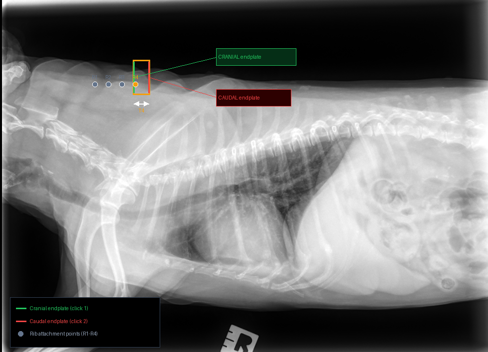
2
Mark T4 Vertebra — 2 Clicks
Zoom into the TOP edge of the image (the spine). Count 4 vertebrae from the left (head) side. Click the cranial then caudal face of that vertebra.
👆
Click the CRANIAL endplate of T4 (head-side face — left side of the vertebra)
● Click 1 = Cranial endplate (head side)
● Click 2 = Caudal endplate (tail side)
Spine at TOP, head on LEFT
100%
|
Where is T4?
1. Look at the very top edge of the image — that bright white segmented row is the spine.
2. Starting from the left (head) side, count 4 vertebral bodies along the spine.
3. The 4th one is T4. It lines up roughly with where the 4th rib attaches.
Each vertebral body is a small rectangular block ~5–10 mm wide. Zoom in to see them clearly.
Sample: T4 on lateral view
Tip: Zoom in 3–4× on the TOP of the image, pan to find the 4th vertebra, then click the left face (cranial) and right face (caudal) of that one vertebral body only.
Step 2
Mark T4 Vertebra
3
Draw Cardiac Axes — 4 Clicks
Click apex then base of the heart (Long Axis), then two ends of the widest width (Short Axis).
👆
Click the APEX of the heart (bottom-most tip of the cardiac silhouette)
● Click 1 = Apex ● Click 2 = Base → Long Axis (L)
● Click 3 = Widest midpoint ● Click 4 = Width → Short Axis (S) auto-locked 90° to L
100%
|
Step 3
Draw Cardiac Axes
Step 4
Mark VLAS Line
VLAS = distance from the carina (tracheal bifurcation) to the bottom of the left atrium, divided by T4.
Click 2 points: 1 Carina → 2 Bottom of Left Atrium
? Reference Diagram
Click 2 points: 1 Carina → 2 Bottom of Left Atrium
Click the CARINA (tracheal bifurcation point)
100%
|
Step 4
Mark VLAS Line
Step 5
Mark Tracheal Line
Draw a line along the distal trachea (last 3–4 cm before carina). The angle this makes with the spine indicates if the left atrium is pushing the trachea upward.
1 Trachea start → 2 Carina end
1 Trachea start → 2 Carina end
Click the start of the distal trachea
100%
|
Step 6
Mark CVC Width
Draw a line across the caudal vena cava (CVC) — the large vessel entering the right side of the heart from below. Measure its widest diameter.
1 One wall of CVC → 2 Opposite wall
1 One wall of CVC → 2 Opposite wall
Click one wall of the CVC
100%
|
Step 7
Mark Aorta Width
Draw a line across the aorta at the same level as the CVC. Used as the reference denominator for the CVC:Ao ratio (normal CVC ≤ aorta width).
1 One wall of aorta → 2 Opposite wall
1 One wall of aorta → 2 Opposite wall
Click one wall of the aorta
100%
|
Step 8
Mark RLAD
RLAD = Left Atrial dimension measured perpendicular to the spine at the level of the carina — the dorsoventral height of the left atrium.
1 Dorsal LA border → 2 Ventral LA border
1 Dorsal LA border → 2 Ventral LA border
Click the dorsal (upper) border of the left atrium
100%
|
Image Extraction Preview
Analyzing radiograph with Claude AI — please wait…
Diagnostic Report
🩻
Awaiting radiograph…
🧠 AI Clinical Triage Steps
❤️ Vertebral Heart Score (VHS)
🫀 Vertebral Left Atrial Size (VLAS)
⚠️ AI-assisted analysis only. A licensed veterinarian must make all clinical decisions.
This summary was prepared to help you understand your pet's X-ray results. Your veterinarian will discuss findings and next steps with you in detail.